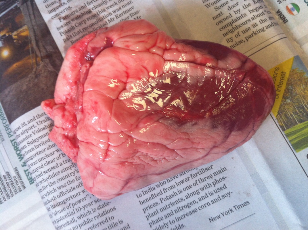
Pictures of the heart we disected.
Heart Dissection
External Examination of the Heart
Examine the exterior of the heart. Refer to the diagrams above to help you.
1. Describe the appearance of the heart. What does it look like? How does it feel? Are there any features you can describe?
The heart was very tough, rock hard, cold and slimy. It was pink-red-purple, in colour and you could see a white fatty substance, you could also see the capillaries. The inside was very fleshy.
3. Find the blood vessels on the surface of the heart muscle. These are the coronary arteries. They carry nutrients and oxygen to the heart muscle
Describe what this artery looks like.
It looked lighter in colour than the rest of the heart, it looked very red.
What do you think would happen if this artery was blocked by a clot?
If the coronary artery was blocked, blood and oxygen flow would be reduced to the heart. Reduced oxygen to the heart can cause a condition called angina.
4. How do you know which is the left and right ride of the heart?
The left side of the heat would be thicker and have more muscles.
5. Have a feel of the thickness of the heart muscle at the top and bottom of the heart. Describe the following features;
a) The thickness of the muscles at the top of the heart.
Firm, doesn't give when pressure is applied.
b) The thickness of the muscles at the bottom of the heart.
Not as firm as the top of the heart, but still tough, though its easier to manipulate.
c) The amount of fat surrounding the heart.
Really hard at the top but at the bottom it not as tough and more flexible, its softer at the back and harder than the muscles.
d) Any major vessels entering and exiting the heart.
Aorta- tougher muscle, thick wall of muscle between the vena cava and the aorta.
6.
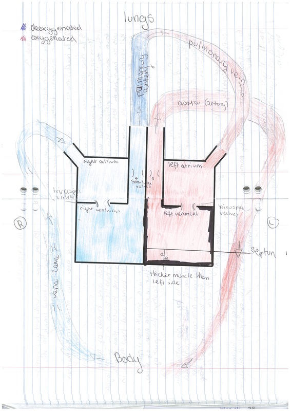
Deoxygenated blood leaves the right atrium in an artery and travels to the lungs. Here, the blood collects oxygen, so it is now oxygenated. The blood travels back to the heart via a vein.
7. Find the aorta that carries the blood away from the left ventricle of the heart.
a) Describe the thickness of this vessel. Why do you think it needs to be so thick?
The vessel is very strong and very thick. It needs to be very thick as its under a lot of pressure, because of this it also needs to be very elastic.
b) Where is it taking blood to?
It is taking oxygenated blood to the rest of the body.
8. Find the vena cava. This is the vein that returns blood from the body.
a) Compare the thickness of the vena cava to the aorta. Why do you think it is different?
The vena cave looks thinner than the aorta and also looks bigger.
b) What part of the heart does the vena cava go back in to?
The right atrium.
c) Remember what you observed when you observed the water flowing through the heart. The water went into the vena cava and into the heart. Which blood vessel did the water come out of the heart from?
The water came out of the pulmonary artery.
9. When the water was flowing into the pulmonary vein, which vessel did it come out of?
The water came out the aorta.
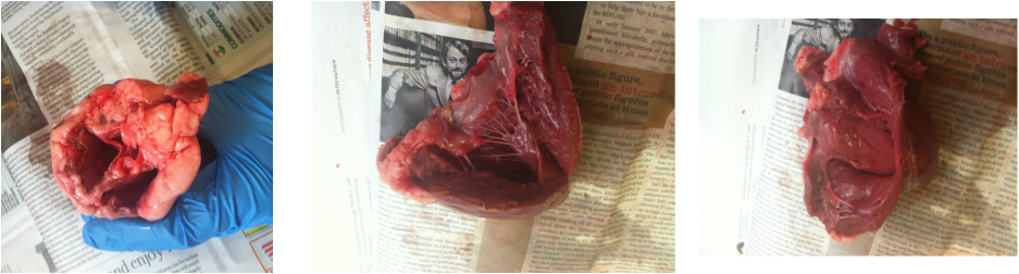
Firstly cut open the LEFT VENTRICLE following the lines on the diagram above.
1. Describe what you see inside the left side of the heart.
A pocket of muscle with strands of tissue or fat attached.
2. Observe any valves you see. What do you think their job would be?
3. Cut the aorta. Describe how it appears and how it feels and any other features
Very hard to cut, very strong.
Examine the exterior of the heart. Refer to the diagrams above to help you.
1. Describe the appearance of the heart. What does it look like? How does it feel? Are there any features you can describe?
The heart was very tough, rock hard, cold and slimy. It was pink-red-purple, in colour and you could see a white fatty substance, you could also see the capillaries. The inside was very fleshy.
3. Find the blood vessels on the surface of the heart muscle. These are the coronary arteries. They carry nutrients and oxygen to the heart muscle
Describe what this artery looks like.
It looked lighter in colour than the rest of the heart, it looked very red.
What do you think would happen if this artery was blocked by a clot?
If the coronary artery was blocked, blood and oxygen flow would be reduced to the heart. Reduced oxygen to the heart can cause a condition called angina.
4. How do you know which is the left and right ride of the heart?
The left side of the heat would be thicker and have more muscles.
5. Have a feel of the thickness of the heart muscle at the top and bottom of the heart. Describe the following features;
a) The thickness of the muscles at the top of the heart.
Firm, doesn't give when pressure is applied.
b) The thickness of the muscles at the bottom of the heart.
Not as firm as the top of the heart, but still tough, though its easier to manipulate.
c) The amount of fat surrounding the heart.
Really hard at the top but at the bottom it not as tough and more flexible, its softer at the back and harder than the muscles.
d) Any major vessels entering and exiting the heart.
Aorta- tougher muscle, thick wall of muscle between the vena cava and the aorta.
6.
Deoxygenated blood leaves the right atrium in an artery and travels to the lungs. Here, the blood collects oxygen, so it is now oxygenated. The blood travels back to the heart via a vein.
7. Find the aorta that carries the blood away from the left ventricle of the heart.
a) Describe the thickness of this vessel. Why do you think it needs to be so thick?
The vessel is very strong and very thick. It needs to be very thick as its under a lot of pressure, because of this it also needs to be very elastic.
b) Where is it taking blood to?
It is taking oxygenated blood to the rest of the body.
8. Find the vena cava. This is the vein that returns blood from the body.
a) Compare the thickness of the vena cava to the aorta. Why do you think it is different?
The vena cave looks thinner than the aorta and also looks bigger.
b) What part of the heart does the vena cava go back in to?
The right atrium.
c) Remember what you observed when you observed the water flowing through the heart. The water went into the vena cava and into the heart. Which blood vessel did the water come out of the heart from?
The water came out of the pulmonary artery.
9. When the water was flowing into the pulmonary vein, which vessel did it come out of?
The water came out the aorta.
Firstly cut open the LEFT VENTRICLE following the lines on the diagram above.
1. Describe what you see inside the left side of the heart.
A pocket of muscle with strands of tissue or fat attached.
2. Observe any valves you see. What do you think their job would be?
3. Cut the aorta. Describe how it appears and how it feels and any other features
Very hard to cut, very strong.


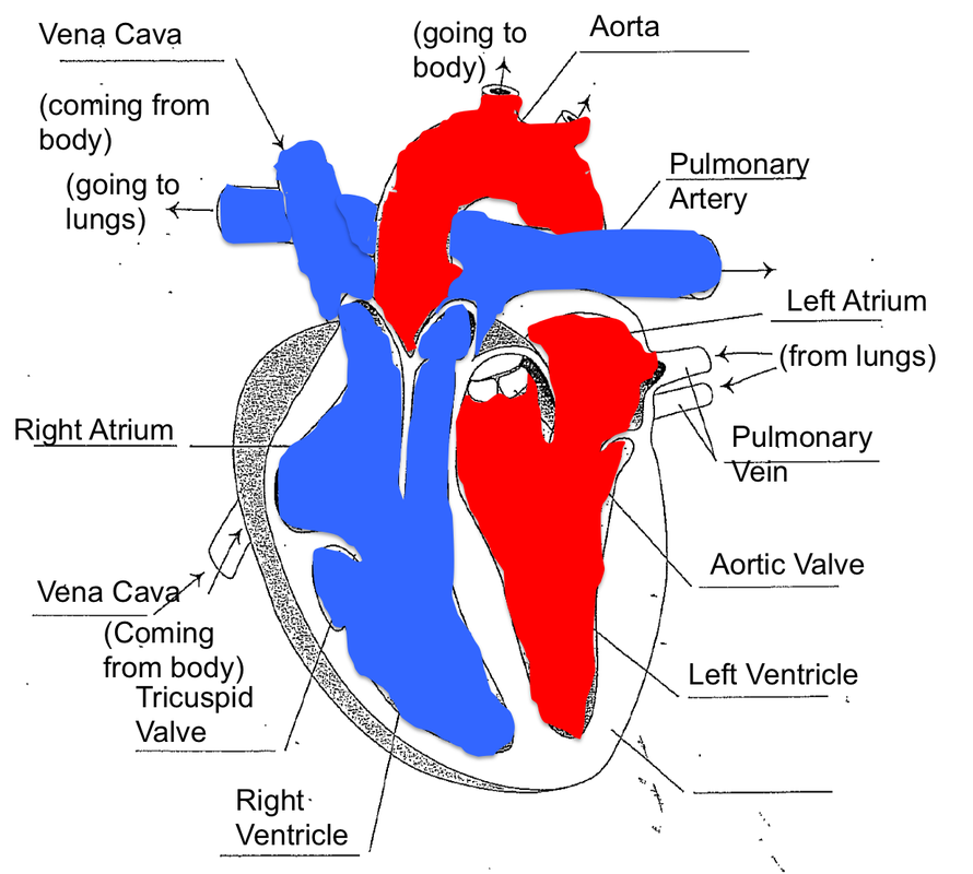
An annotated picture of the heart, explaining the names of different parts
A box diagram of the heart, this shows all the different parts of the heart.
